CELL_STRUCTURE
CELL
Cell is the unit of
structure and function. They are the building blocks of an organism.
Irrespective of the nature of organisms (plant or animal) they are either made
up of single cell or many cells, the former is called unicellular and later is
called multicellular organisms; in the later cells are differentiated into
various kinds and they are grouped into tissues, which perform special and
special function.


Example meristematic cells perform repeated
cell divisions, phloem cells- conduct food material, sclerenchyma- mechanical,
xylem- conduction of water and mineral salts and so on. Nevertheless, all these
different types of cells are derived from the same embryonic cells. The
development of various cell types from a single cell is determined and
regulated by a process called differentiation, which in turn is controlled by
differential concentrations of plant hormones. Added to this, organ
differentiation is another fascinating aspect of development. All these
processes are regulated by differential gene regulation in response to
environmental stimulus and phytohormones.
Cellular composition:
All cells are made up
of a semi viscous fluid called protoplasm, which is considered as the physical
basis of life, for it controls all biochemical reactions of the cell. In fact
it is the microcosm of life with many secrets not known to us. The protoplasm
is colloidal in nature, because many cell colloidal sized structures and
macromolecules are suspended in it. It also exhibits sol and gel properties.
The granular nature of the protoplasm is due to the presence of many tiny
organelles. Vacuoles of various types are also found, but in plant cells when
it is matured, a large central vacuole is present and it is separated from the
rest of the protoplasm by a single unit membrane called tonoplast. The fluid
present is called cell sap. There are many common features between plant and
animal cells; the former is distinguished by the presence of distinct cell wall
and plastids, which are totally absent in animal cells. However centrosomes are
invariably present in animal cells and rarely in plant cells with exception of
some lower plant unicellular algae like Chlamydomonas.
Chemical composition
When cells are
subjected to chemical analysis the following compounds are found (approximate
value).
Compound Animal plant
cell
Carbohydrates
20% 30%
Proteins 45% 40%
Lipids 30% 25%
RNA 1% 0.4%
DNA 0.2% 0.4%
Inorganic
& others 3.8% 3.6%
Carbohydrates: These are organic
compounds consisting of C, H and O. The basic structural components of
carbohydrates are monosaccharide sugars consisting of 3 to 7 carbons. Example:
Glucose (6c), Fructose (6c), Erythrose (4c), Xylose (5c), etc. such monomers by
undergoing polymerization develop into long chained polysaccharides, such as
cellulose, cellobioses, starch and glycogen (animal starch). Cellulose is made
up of glucose units linked by β1>4 linkages and it is an important
component of cell wall. Similarly starch is also a polymer of α1-4 linked glucose
units and it is the main source of energy for all living cells.
Proteins: proteins are the most
important organic components of cells, for they act as structural as well as
functional molecules. With out proteins life cannot exist, though DNA is the
genetic material it is proteins that make it or break it; DNA is master library
with information, that cannot be changed. They are made up of basic building
blocks called amino acids. The polymers of amino acid residues are called
polypeptides (proteins) which exhibit different structural conformation
(shape). The 3-D shapes are specific and characteristic for a particular
protein, thus they exhibit specific structure and function. Exemple:
contractile proteins (muscles), transport proteins (Hemoglobin), enzyme
proteins, hormonal proteins (insulin), antibody proteins IgG etc. Almost all biochemical
activities, including growth and development are controlled by proteins,
without which cell ceases to live.
Lipids: Fatty acids and their
derivatives are very important for two reasons, firstly lipids like lecithin,
phosphotidyl ethanolamine, sphingolipids, glycolipids, steroids and others are
part of the cellular membranes; thus they contribute to the structure of the
membranes. Secondly lipids also act as food reserve and provide energy by
oxidation.
Nucleic
acids: These are the polymers of nucleotides, consisting of
nitrogenous bases, phosphates and pentose sugars. There are two types of
nucleic acid – 1) Deoxy-ribose nucleic acid (DNA), 2) Ribose nucleic acid
(RNA). DNA is mostly found in chromosomes, it is the repository of genetic
information and it provides information for the synthesis of proteins. The
chemical composition, structure and function will be discussed in the chapter
elsewhere.
Inorganic/Organic
factors : Many inorganic metals Fe, Mg2+, Mn2+, Mo, ca2+ etc. are
very important, for they are either the integral part of some organic molecules
or act as co-factor in enzymatic activities.
Certain organic
factors like important for cellular metabolism, because many of the vitamins
act as co-enzymes; these are required for enzymatic activities, without which
cellular processes come to stand still.
Size and shape: The size of cells varies from 10 micron to
many centimeters in length. For example cotton fibers are several mm in length
The shape ranges from spherical, isodiametric, and hexagonal to tubular. This
is genetically predetermined to perform different functions.
Cell structure:
When the cell is
observed through light microscopes, which may have the maximum resolution of
about 2000 times, very few details can be made out. On the other hand, if
sections of the cells are observed through electron microscope, which has a
resolution power ranging from 50,000 to 150,000 times enlargement, even smaller
structures stand out clearly. Inspite of high resolution, it is not possible to
make out all the structural details.
The following structures
are found in the cell, 1) Cell wall, 2) Plasma membrane, 3) Nucleus, 4)
Plastids, 5) Mitochondria, 6) Golgi complex, 7) Ribosomes, 8) Cytoskeleton, 9) Micro
bodies, 10) Centrosomes,11) endoplasmic reticulum, 12) central vacuole and 13)
Non living cell inclusions.
Cell wall: Only plant cells and
bacterial cells posses a protective structure as the cell wall outside the
plasma membrane. Bacterial cell wall is firmly adpressed to the underlying
plasma membrane.
Its
chemical
composition and structure is more complex. However, it is basically made
up of
long polymers of glucosamine (NAG) and muramic acids (NAM), which in
turn are cross linked by short-pentamer oligopeptides; thus they form a
mat
like structural layers around the plasma membrane. Hundreds of such
layers are
deposited one above the other to form a very tough wall. Many bacterial
cells
produce a mucilaginous pectose layers outside the cell wall and this
layer is
called the capsule. But many bacterial cells do contain another lipid
layer
studded with proteins, oligosaccharides and glycol and proteoglycans.
But higher plants have
cell walls mainly made up of cellulose fibres. Addition to these, pectins, hemicelluloses
and lignin are also deposited on primary cellulose layer; the thickening is only
at the later stages of development.
With proper staining,
if a group of cells are observed, the cells appear to be held together by a
kind of cementing material called middle lamella. It is made up if calcium
pectate. Next to calcium pectate layer, the cell wall that is laid later is
differentiated into 2 or 3 distinct layers. They are primary wall, secondary
wall and tertiary wall.
The primary wall is
the first true cell wall to be deposited and it is purely made up of cellulose.
With the development of the cell, the additional layers are deposited, called
secondary and tertiary wall layers, like in the case xylem and sclerenchyma.
Certain areas in the cell walls remain without thickening and these are called
pit areas, through which fine protoplasmic strands transverse across between
two cells and provide a continuum to protoplasm; they are called plasmodesmata.
The primary cell wall,
which is the first wall layer to be deposited, is made up of cellulose fibres.
These fibres are considerably longer and deposited layers after layers;
oriented longitudinally, transversely or obliquely. The deposition and the
orientation of these layers are aided by microtubules that are found on the
inner face of the plasma membrane.
Each cellulose fiber is
made up of 8000 to 120,800 D- Glucose units, which are linked to each other by glycosidic
bonds to form a long chain of glucose units, which show helical conformation.
Hundreds of such cellulose threads are grouped into a bundle called micelles;
these in turn are aggregated into micro fibrils which by further aggregation
develop in to macro fibrils.
Such micro/macro
fibrils are deposited regularly either longitudinally or transversely to form
uniform layers. These fibres are embedded in a matrix made up of pectate
substances and hemi cellulose materials like polyxylose and others.
Primary cell wall Secondary cell wall
Primarily cellulose Few
layers of cellulose
Hemicelluloses Xylose,
mannose
Pectic Complex
lignins
Elastic Non-elastic
Laid on middle
lamellae Laid over primary wall
1-3 micron thick >5-10
micron thick
Cell membranes
These are the most
important structures of the cell, for they are responsible for protecting and
separating the protoplasm from the external environment.
It helps in selective uptake and transport
of ions, provides surface area for many biochemical signal transduction
reaction and also helps in specialized functions. In fact most of the cell
organelles are bounded by membranes. Notwithstanding this, a large number of
functional molecules are integrated into these membranes.
Chemical composition
Almost all membranes are made up of
proteins and lipids. The ratio between proteins and lipids may vary in
different membranes, but generally it is equal. It is not uncommon to see some
carbohydrates as glycoproteins associated with membrane on the outer surface of
plasma membranes. Proteins found in membrane are not of the same kind, but
differ in their structure, chemical composition and function. Some of them are
large and many of them may be smaller. However, most of the proteins are
globular in nature having either hydrophilic or hydrophobic or both the
characters. Lipids, on the other hand are of various kinds. The common lipids
found in the membranes are phosphotidyl choline (Lecithin), phosphotidyl
ethanolamine, Phospholipids glycerol, cholesterol, etc. some of the
phospholipids exhibit hydrophilic groups at one end and hydrophobic at the
other end. Nevertheless, the composition of lipids varies from membrane to
membrane, for they have different functions to perform.
Membrane structure
The structural organization
of various compounds with in the membrane was an enigma for a long time. With
the advancement of biological techniques, it has been found that the membranes
exhibit fluid-mosaic structure (Singer and Nicholson 1972). Basically lipids form
bilayers by organizing hydrophobic layers facing each other and the charged
region out side. Proteins, depending upon the charged nature, some are found on
the surface and some are embedded. But proteins & lipids of various kinds
are oriented towards each other in such a way, they exhibit semi-solid
(crystalline) and semi fluid properties. The arrangement of lipids and proteins
is of mosaic pattern, where many proteins are half buried in the lipid bilayers
and some traverse the entire cross section of the lipid layer in such a way a
part it is buried in the core and some are located at the peripheral surface.
The structural and chemical heterogeneity is the hall mark of these membrane
structures. This model explains various biological phenomena observed in most
of the biological systems.
Various membranes
found in the cell, like plasma membrane, endoplasmic reticulum; organelle
membranes exhibit basically the same structural pattern, but vary in their
lipid and protein composition. Some of them are single unit membranes (plasma
membrane, tonoplast, Lysosomes and peroxisomes) and some have double membrane
systems (endoplasmic reticulum, nuclear membrane, golgi membranes chloroplast
membrane and mitochondrial membrane). However they show differences in their
chemical composition structural features.
Plasma membrane
This is the outer most
membrane of the cell, within which all protoplasmic structures are included. In
plants, cell wall acts as an additional protective layer, but in animal cell, plasma
membrane itself is the bounding membrane.
This membrane is in
immediate contact with the external environment, and performs various functions
like osmosis, selective absorption of various mineral nutrients, accommodates
innumerable signal transducing receptors for various stimuli (electrical, light
mechanical and chemical) and invariably exhibits dynamic properties. Proteins
found in the plasma membrane are vectorial (directional) arranged. At some
places, the plasma membrane shows inward projections and they are in continuity
with the endoplasmic reticulum. Plasma membrane is also the site for
pinocytosis and phagocytosis. Thus the plasma membrane exhibits unique but varied
properties of its own. It exhibits dynamic fluidity never to be constant and
stagnant.
Endoplasmic Reticulum:
It is a labyrinthine
net work of double membrane sheets. They are found in all living cells with the
exceptions of mature erythrocytes and cells of bacteria. Endoplasmic reticulum
(ER) occupies more than 50 to 90% of total cell volume. It is in contact with
the outer plasma lemma and the outer nuclear membrane. ER is made up of two
single unit membranes folded to form adpressed sheets, which enclose a channel
or cisternae. ER is extensively branched, thus the surface area for reactions is
enormously increased.
On the basis of
presence or absence of ribosomes on the surface, two classes of ER can be
recognized
1)
Smooth ER (SER) is without ribosomes, 2) Rough ER (RER) has innumerable
ribosomes on its outer surface. These membranes are highly mobile and they
undergo rapid flux, changing from SER to RER and RER to SER. Added to this, the
entire ER exhibit continuous sweeping movements which help in the distribution
of cellular components in the matrix. The ER membranes are supported by
microtubule skeletal network. These membranes are highly dynamic and show rapid
turnover.
Functionally ER
exhibits various activities like synthesis of proteins, mechanical support for
the fluid protoplasm, synthesis and storage of lipids, synthesis and storage
and transport of proteins to different destination through golgi membranes.
Some of the functions are detoxification, transport of various cellular
components, formation of micro bodies, formation of secretory vesicles, cell
plate and others. The above mentioned functions indicate that the ER is at the
heart of various cellular activities. Furthermore, during the development of
nucleus and other cell organelles like, chloroplast, mitochondria, golgi complex,
micro bodies, Lysosomes etc. ER provides membrane fractions to them. Exchange
of membrane components among them is pervasive and common.
Golgi bodies
A group of membrane
cisternae, discovered by Camillo Golgi in 1890, are called as Golgi bodies.
They are present in all cells except bacterial and blue-green algal cells.
These structures vary in number from cell type to cell type. In secretory
tissues like thyroid and liver they are present in large numbers than in other
type of cells. They are abundant in secretory surfaces like stigma of the
pistils.
Golgi bodies are made
up of a group of stacked membrane cisternae. Some times these membranes
structures show extensive reticular network. Generally the distal ends of
double membranes are dilated into vesicles and some of the vesicles are in the
process of pinching off. An interesting feature is that the Golgi complex is
surrounded by ER and at some places they look like in continuity with ER, but
these proximal membranes of ER are free from ribosomes called SER (smooth ER).
The stacked Golgi
membranes have two faces, i.e. formation face is called cis face and maturation
face as trans face. The formation face has convex surface and has a number of
small vesicles pinched off from SER. In fact certain proteins synthesized on
the RER, enter into the lumen of ER and hence they are transported in the lumen
towards the transitional membranes which are in close association with Golgi
complex and then the protein containing ER membranes pinch off as vesicles.
These in turn fuse with one another or with golgi cis membranes and develop
into membranous sacs. Within these membranous sacs, proteins and such products
get further modified. Later such products are sorted out and get enclosed and
pinched off in the form of vesicles from maturation face called Trans surface of
the Golgi complex. Similarly many secretary products that are synthesized on
RER and transported into Golgi complex, later the matured products get enclosed
in vesicles and budded off. The golgi derived vesicles are loaded with
proteins that are specifically targeted to various destinations.
Thus, Golgi bodies
perform various complex processes, like glocolysation of proteins, synthesis of
cell wall polysaccharides, maturation of zymogen granules, formation of primary
lysosomes, secretion of lipid bodies, acrosome formation, neural secretion
etc. Nevertheless, the participation of ER is every essential in the function
of Golgi membranes. Golgi membranes are associated with transport proteins and
they are responsible for transportation from one site to the other. In all the
above mentioned processes, packing, maturation and secretion of specific
substances are the most important events of Golgi functions. The golgi complex
is at the heart of membrane flow.
Lysosomes
Though these organelles
were noticed in 1949, later it was de Duve who coined the term Lysosomes for
such dense bodies. Lysosomes are often called suicidal bags, misnomer, for
they are capable of digesting various cellular structures and digest every
thing, if they are damage or made to break open to release the contents.
Without any exception
all eukaryotic organisms contain these bodies in their cells. These are found
in various sizes. Lysosomes are bounded by a single unit membrane and enclose
a group of hydrolytic enzymes. Paradoxically, the surrounding membrane is not
digested by the enclosed hydrolytic enzyme; this is probably due to special
modifications of the membrane and lysosomal fluid which is more acidic. The
intactness of the membranes is mainly dependent upon certain membrane
stabilizers like cholesterol, cortisones, cortisols, vitamin E, antihistamines,
heparin etc. On the other hand, substances like Vitamin A, Vitamin B, Vitamin
K, B-estradiol, testosterone and digitonin labalize and cause leakiness.
Sometimes at higher doses, the membrane may completely disperse and all the
lysosomal contents may be released. As a result the cell may be completely
digested.
Lysosomes are
important cell organelles in digesting various macromolecules like
carbohydrates, proteins, fats, DNA, RNA and others. The breakdown of these
molecules during various stages of development and metabolism is governed by
the controlled release of these enzymes.
The origin and
development of lysosome itself is controlled by many environmental factors.
For example, when an organism is starved of food, lysosomal number increases
tremendously. This is in order to degrade whatever food material that is
available in the cell. Similarly, when yeast cells are subjected to anaerobic
conditions or starved food, within minutes, Lysosomes increase in number and
actively; they chew up all the available materials including mitochondria. In
the case of germinating castor seeds or maize grains the increased lysosomal
activity helps in the degradation of fats and starch into simple molecules for
the growing seedlings. The number of enzymes and the kinds of enzymes found in
each of lysosomes is not same or constant. The contents vary depending upon
the tissue and the metabolic status of that tissue. Commonly available
lysosomal enzymes are nucleases, phosphotases, lipases, proteases, glycosidases
and sulfatases. Even some of the condemned proteins are taken in through LAmP
proteins on its membrane surface and digest the same. Many of the lysosomal enzymes
are released in a regulated way and they have definite pH optima for their peak
activity. Lysosomes and some trans golgi vesicles and incoming endosomes form a
kind of network which provide material for its growth and maintenance.
Curiously enough,
lysosomes take their origin from Golgi bodies. Lysosomes enzymes are
synthesized on RER: then they are transported to smooth transitional vesicles.
Afterwards they are integrated into Golgi sacs at the formation face. After
undergoing modifications and packing, they are then sorted into vesicles, which
are budded off from maturation face of the Golgi complex. The vesicles
containing lysosomal enzyme are marked by the addition mannose6-phosphate, such
vesicles ultimately dock with lysosomes via late endosomes or directly. These
may further fuse with one another to form larger structures or they may fuse endosomes
or and with phagocytotic vesicles (phagosomes) to form secondary lysosomes,
where the engulfed substances are digested and the products are resorbed into
cytoplasm. The undigested materials are removed by defecation. Many a times,
the lysosomes move towards the plasma membrane and unload all their contents,
thus they cause extracellular digestion. Lysosomes also play a significant
role in the acrosome formation (cap structure) of spermatozoid. The presence of
such cap structures in sperms help in their penetration through the tough
cortical walls of the egg cells.
Lysosomes are also
known to play a very important role in metamorphosis of amphibians and
insects. For example, during the transformation of tadpole into adult frog;
the long tail of the tadpole gets digested by the lysosomal activity, the process
called resorption. Recent investigations have further shown, that the
increased activity of lysosomes causes severe destruction of tissues, probably
of lysosomes cause severe destruction of tissues, probable break down of
chromosomes leading to abnormality; perhaps even cancer may be induced.
Another instance of lysosome induced disease is Rheumatoid arthritis (Joint
pain). Thus lysosomes play a significant role in the metabolism and
development of an organism. One can find various lysosomal based human
diseases.
Microbodies
These are spherical,
electron dense, granular bodies. They are bounded by a single unit membrane.
Such structures are found in both animal and plant cells. Their number varies
from 50 to 100 per cell; their number can increase or decrease based on the
requirement. Germinating seed cells show maximum numbers; often they form a
link between chloroplast and mitochondria, where peroxisomes are involved in the
detoxification of oxygen free radicals by catalase or peroxidase activity.
There are two types of
micro bodies and they are characterized by their functions viz., 1) peroxisome,
2) glyoxysomes; the former exhibit peroxidase activity and the later show
glyoxalate activity. Such bodies are found in various tissues like liver,
kidney, intestine, brain, lung epithelial cell, testis, brain, adipose, and
photosynthetic cells of green plants.
In C3 plant cells (Calvin
plants) they are closely associated with mitochondria and chloroplasts. They
are responsible for photorespiration. This process severely results in the
depletion of photosynthetic products. However, they are not that active in C4
plants. Nevertheless in C3 plants both Glyoxysomes and Peroxisome act
co-operatively utilizing Ru-DP glyoxalate and breaking it down to glycollate,
which is then metabolized by peroxisome. On the other hand in animal tissues
like liver and other cells, various substances like urea, amino acids, lactic
acid etc. are oxidized by peroxisome to H2O2. In this process oxygen is
utilized. As H2O2 as peroxide is fatal to living cells, it is salvaged or
removed by superoxide dismutase (SOD) reaction and oxidation to water by
utilizing substrates like ethyl alcohol, methyl alcohol, nitrates, etc. The
presence of these structures and their function is fascinating, for their exact
role is not clearly understood. They are also implicated in thermoregulation
of certain organisms.
Ribosomes:
Ribosomes are
ultramicroscopic particles, first observed by Palade. Though they are
submicroscopic in size, they are extremely important, for they are responsible
for the synthesis of proteins.
Ribosomes are found in
millions, in eukaryotic cytoplasm, but in bacteria, ribosomal count is approx.
20000. They are also found in organelles like chloroplasts and mitochondria.
These structures are either roughly spherical or ovoid in shape. They are very
stable and can remain functional at least 120 days or so. Basing on the
molecular weight (determined by equilibrium density ultra centrifugation) they
have been broadly classified into two types, i.e. 80 S and 70 S types. The
S-values of the slightly vary among the organelle ribosomes. Ribosomes of 80s
type are exclusively found in eukaryotic organisms.
But 70 s ribosomes are
restricted to prokaryotic organisms like bacteria and blue-green algae. Still
smaller ribosomes are found in mitochondria and chloroplasts, but their sizes
vary.
When functional
ribosomes are subjected to Mg2+ depletion, they separate into large and small
subunits. If the concentration of Mg2+ is increased the free subunits
reassemble into functional units. The concentration of Mg2+, if further
increased, ribosomes aggregate into dimmers, tetramers or octamers, thus one
can precipitate ribosomes and collect them by centrifugation.
The smaller subunit
under electron microscope exhibits a shape of elongated cucumber with an
indentation and a twist. But the larger subunit shows a shape of a mother in
sitting position with the knees upright, having the baby on her lap. Here the
‘Baby’ is equivalent to a smaller submit (note this purely my imaginary
explanation not found in any text books). The larger subunit has a grove in
the middle through which the nascent polypeptide traverses and exits.
Ribosomes found in the
cytoplasm exist either in membrane bound state or free state. The ratio
between these varies from cell to cell type and depends upon the functional
state of the cell. But in bacteria most of the ribosomes exist in free state.
Chemical analysis of
these structures indicates that they are made up of proteins and ribose nucleic
acids (rRNA) roughly in equal ratios. The proteins of larger subunit of
prokaryotic bacteria consist of 34 subunits called L1 to L34 and the smaller
subunit has 21 factions, s1 to s21. Each one of these proteins is unique with
the exception of two proteins i.e. S 20 and S 26 which are present in both the
ribosomal units.
Ribosomal RNA exists
in three or four different sizes which are expressed in Svedberg units (S) as
shown in the table below.
The RNAs present in
ribosomes show various sizes, and most of them are rich in Guanosine and
Cytosine nucleotides. In prokaryotes rRNAs coded for by seven operons as
precursor containing 16s, 23s and 5s RNA. Such precursors also contain tRNA
blocks tucked in them. The genes responsible for the synthesis in eukaryotic
cells are found in multiple copies i.e. of 200-500 organized as tandem repeats.
Further more rRNA genes are exclusively found in secondary constrictions or
nucleolar organizer region of the chromosomes, in humans they are located on
chromosomes 13, 14, 15 21 and 22. The rRNA gene contain 28S, 5.8sRNA and 28sRNA
gene, but the 5 S RNA genes are found elsewhere and spread over other
chromosomes. These RNAs are transcribed on rRNA genes as larger precursor molecules
(45 S RNA) and then they are cut and processed into smaller molecules such as
28 S, 18 S and 5.8 S RNAs. Notwithstanding this, the ribosomal RNA genes in the
case of Frog oocytes are amplified 1000 fold. Enormous number of ribosomes are
synthesized and accumulated in the eggs. Another interesting aspect of
ribosomes is that, various ribosomes proteins are coded for by the chromosomal
DNA other than the nucleolar DNA. They are synthesized in cytoplasm and they
are transported into nucleolar region of the nucleus. There, the rRNA and
ribosomal proteins associate to form functional subunits of ribosomes in an hierarchical
fashion. Later they are transported to cytoplasm through nuclear pore complexes
to perform protein synthesis.
However, mitochondria and
chloroplast employ a different mechanism for the assembly of ribosomal
components. Organelles synthesize their own rRNAs and get imported riboproteins
from cytoplasm and assemble functional ribosomes which more or less similar to
prokaryotic ribosomes in size and function. Thei activity can be inhibited by
chloramphenicol.
The quintessential
function of ribosomes is to act as dynamic machinery for the synthesis of
proteins. This is achieved by the association of ribosomes with messenger RNA
(m.RNA) and amino acid loaded transfer RNAs (aat.RNA). Many ribosomes may be
associated with a single m.RNA and such a cluster is called polysome. These
polysomes may be free from membrane, or membrane bound.
Plastids:
Plastids are very
important cell organelles found mostly in plant cells. They are mainly
responsible for photosynthesis.
Green color of plants
is due to the presence of green pigments in plastids. Other colors found in
various structures like leaf and flowers are due to the presence of other colored
pigments in the plastids (other than green). Basing on the presence or absence
of pigments, plastids have been broadly classified into colorless plastids (Leucoplasts)
and color plastids (Chromoplasts). If the plant tissue containing Leucoplasts
are exposed to sunlight, they may turn into chromoplasts. On the contrary, if
plants with chromoplasts are kept in dark for sufficient time, the chromoplasts
will be transformed into colorless plastids.
Leucoplasts are
generally found in roots and inner tissues, which do not receive sunlight.
Such plastids normally store different kinds of food materials like starch,
proteins and oils; basing on this they are called Amyloplasts (store starch),
Proteinoplasts (proteins) and Elaioplasts (oil). Such structures are also
found in abundance in various storage organs like fruits, tubers and oil seeds.
Chromoplasts, on the
other hand depending upon the dominant pigments present, are classified into
chloroplasts (green) Rhodoplasts (Red), Fucoplasts (brown), Xanthoplasts (Yellow)
and so on. Majority of terrestrial plants and aquatic plants like green algae
contain chloroplasts. It is important to note that plants found in ocean which
account for the major part of vegetation on this planet, contain different
kinds of chromoplasts and also green plastids. While eukaryotic plants (higher
plants) contain well developed plastids, plants like photosynthetic bacteria
and blue green algae (prokaryotic) do not possess such plastids, but still they
perform photosynthetic functions because the photosynthetic pigments are either
found organized in the form of stacks of membranes or as vesicular structures
called chromatophores.
Number: The number of
plastids, particularly in green plants, varies from one to hundred or more.
Plants like Chlamydomonas have only one chloroplast; spirogyra has two spirally
coiled chloroplasts and in higher green plants like angiosperms the number
varies from 20 to 100 per cell.
Shape: Chloroplasts show
some interesting variation in their shape. This shape is constant for a given
species. For example in Chlamydomonas, it is cup shaped; in spirogyra it is
spirally coiled and ribbon shaped, in Zygnema it is star shaped, in
Hydrodictyon it is reticulate type and in higher plants it is oval or
spherical. Shape and size of chloroplasts is species specific.
Size: It varies from 0.5
micron to 6 micron in diameter but again its fully formed has specific sizes
and species specific.
Structure: Basically most of the
chloroplasts are bounded by two single unit membranes and the space between them
is called periplastid space. Inside the membrane system, chloroplasts are
filled with a highly complex fluid called stromatal matrix or simply called
stroma. Within this fluid, various specialized structures, macro molecules and
enzymes are either suspended state or the fluid shows flux. The structures
found are grana, ribosomes, DNA, RNAs, starch and other various enzymes,
coenzymes required for carbohydrate and fatty acid synthesis and amino acids.
Grana: These are the most
specialized membrane systems derived from the inner chloroplast membrane. A
single chloroplast may contain 10-30 grana. Each granum is made up of 10-60
circular membranous discs stacked one above the other and such discs are called
thylakoids. The grana are inter-connected by the thylakoid membranous
extensions called inter-granal lamellae or stromal lamellae. However in C4
plants, like sorghum, sugarcane and other tropical grass plants, chloroplasts
show two types of organizations, i.e. the chloroplasts found in the bundle
sheath cells do not possess any differentiated granal structures, but are
filled with diffused stromal membranes, arranged parallel. However,
chloroplasts found in mesophyll cells show granal structures. Chloroplasts in
bundle sheath show centrifugal arrangement, i.e. chloroplasts are oriented
towards the mesophyll cells. Protoplasmic connection is found between
mesophyll cells and bundle sheath cells.
Thylakoid: Thylakoids are the
highly differentiated and say, super specialized circular membranous cisternae,
in which various molecular complexes are embedded and they are responsible for
photochemical reactions. Though the membrane has the same basic components and
organization as that of any other unit membrane, it is the assembly of
photosynthetic pigments and its associated proteins makes it a specialized
membrane system. But photosynthetic pigments like, Chlorophyll-a
Chlorophyll-b, Carotenoids and their associated proteins are grouped in such a
way that they act as units for performing photosynthetic functions. Based on
the chemical composition, size, structure and function, they are classified
into Photosystem I and Photosystem II. Each of these photosynthetic units contain
about 250-300 chlorophyll molecules associated with specific and specialized
proteins. These photosystems appear to be granular in structure of different
dimensions.
If thylakoid membranes
are cut open by freeze-fracture method and observed under electron microscope
they reveal the presence of two types of photosynthetic units of different
dimensions and they are called Quantasomes The larger quantasome, which is
found on the inner surface has a size of 185A^o. They are grouped into an
array of 4 to 6 units. On the other hand the smaller particles are of 110 A0
size and are found arranged on the outer surface of the membrane. The former
particle has been identified as photo system II and the later as photo system
I. The outer surface of the thylakoid membrane i.e. surface A is also studded
with granular particles of different sizes. They have been identified as RuBP
carboxylase and ATP synthetase units. However PS I and PS II are arranged in
such a way they fit into one another like Jig-saw structures. This ‘close and
tight’ fit arrangement helps in the co-coordinated functioning of the
photosystems. But the inter-granal lamella contain only PS I. The chemical
composition of PS I and PS II and their function is discussed in greater detail
in the chapter photosynthesis. There is another important complex called
Cytochrome b6-f complex which acts between these two systems and it is found
both in stromal and thylakoid membranes.
Plastid
Nucleic Acids: Apart from granal structures, various types of nucleic
are found in the stroma. The presence of a circular double helix DNA molecule
of 123 to 200Kbp long is highly significant. This gives the chloroplasts a
status of semi autonomy. Such 10 to 30 circular DNA molecules are found in
each chloroplast. It has been estimated that the information present in DNA is
sufficient to code for ~120 plastid proteins or more. The relationship between
certain segments of DNA and certain proteins of chloroplasts has been
established, however the knowledge about the other regions is not known. Majority
of RNAs like r.RNA, tRNA about 120 species of m.RNA, are coded by chloroplast
DNA. The presence of various types of RNAs and 70s ribosomes, clearly
indicates that chloroplasts are endowed with a machinery, which can synthesize
most of its proteins by itself. Nonetheless, majority of the proteins that are
required for chloroplasts to function are synthesized on cytoplasmic 80S
ribosomes and transported into plastids via chloroplast membrane transport
systems. The interaction and regulation of gene activity between nuclear genes
and plastogenome is an upcoming field of molecular biology, whose scope is
unlimited.
Apart from the above
said structures and molecules, the stromatic fluid also contains an army of enzymes
responsible for carbon pathway, amino acid synthesis, starch synthesis, protein
synthesis, fatty acid synthesis and other enzymes for nucleic acid metabolism.
Among them the RuBP carboxylase is found in very large amounts. In fact, it
has been considered as the most abundant protein in nature, next to only to Tubulins.
This RuBP enzyme protein accounts for more than 50% of the total leaf proteins.
This figure summarizes Light reactions
Biogenesis: Various plastids,
during biogenesis, develop from the pre-existing plastids called proplastid.
They are, in fact, inherited from the maternal side. While the meristematic
cells divide, proplastids too divide by binary fission, just like bacteria, and
get distributed equally or unequally to their daughter cells. When tissues or
cells containing such proplastids are exposed to sunlight, particularly blue
and red lights, they undergo a series of developmental changes. Proplastids
contain a double membrane system. With the onset of light, the plastogenome
gets activated; later certain nuclear genes also get activated required for
plastid development. The future of it depends upon the kind of tissue in which
it is located and other factors, where a plastid can develop in to colored,
other than green, into colored plastids, as found in flowers. Activation
transcriptions not only in the plastid, but also in nuclear genes. This leads
to the transcription and translation required for plastid biogenesis. The
plastid membranes have protein transport system located in a region where one
finds the membranes in close association called attachment points. It is here
cytosol protein transporters are located. It is at this time one can observe
the inner proplastid membrane starts producing inward invaginations in the form
of finger shaped processes. They are loaded with imported proteins. These invagination
in turn start pinching of membranous vesicles in large numbers. These vesicles
then start expanding and later arrange into stacks. Finally they get organized
into granal and intergranal structures. Along with these developments, various
stromatal ingredients are also synthesized and accumulated. The regulated
interaction of nunclear as well as plastid gene activity is a must for the
development of functional chloroplasts. Chloroplasts perform light reactions
where light energy captured, converted into chemical energy and stored in
energy rich bonds of ATP and reducing power NADPH+H. Besides photosynthesis, they
also perform carbohydrate synthesis and fatty acid synthesis and transport.
Mitochondria
Most of the biological
activities are energy dependent and the energy that is required is the chemical
free energy. The provision and production of such energy rich molecules like
ATP, PEP, PGALD, Succinyl Co.A., Acetyl Co.A, NADH2 and such molecules are must
for the sustenance of any cellular function.
Mitochondria are one
of the most important cell organelles, which are endowed with a capacity to
produce energy rich molecules like ATP and NADH+H. In fact, mitochondria are
considered as ‘power plants’ of the cells. Besides producing energy rich
molecules, it also performs fatty acid oxidation to release energy rich
components and other intermediate molecules which are essential for other
metabolic pathways.
Furthermore,
mitochondria are also involved the synthesis of amino acids.
Number,
size and shape: The number of mitochondria per cell ranges from few to
many hundreds or more and depends upon the type of the cell and its metabolic
status. Whenever or wherever, there is a need for higher amount of energy
output, mitochondria are found in large number, example: flight muscles; 1000
or more per cell. On the other hand liver cells contain little less number.
Similarly mesophyll cells of plants, where greater number of chloroplasts are
present, the mitochondria are in negligible numbers, for chloroplasts
themselves satisfy most of the energy needs of the cells. Similarly in
anaerobic yeast cells there are few mitochondria.
The size of mitochondria
is generally between 3 –4 x 0.5 – 10 microns, but under certain pathological
conditions the mitochondria may enlarge considerably, example: diseased thyroid
glands. Normally mitochondria under optical microscopes with a resolution of 200
to 2000 times, appear to be thread like, similar to that of bacilli bacteria,
provided they are stained with specific stains. But other shapes like
spherical, branched and circular are not uncommon. Though mitochondria are
stable, very often they exhibit a rapid turn over, involving changes in the
size, shape and number.
Structure: Mitochondria are
bounded by two single unit membranes. The outer membrane is smooth, but the
outer surface may be studded with some granular structure, probably involved in
glycolytic steps of respiration. The space found in between the outer and
inner membranes is called peri-mitochondrial space. It is filled with a fluid
and rich in cyt.C. The inner membrane is highly folded inwards called
cristae. The number of cristae is not constant and it depends upon the demand
for the energy supply. Nevertheless, the increase in cristae increases the
surface area of reaction within a limited space and volume, an efficient system
indeed.
Cristae: The inner membrane
or cristae is made up of lipids and protein structures. Some of the lipids
present are phosphotidylinositol, phosphotidyl-choline, phosphotidylinositol
and cardiolipin. With regard to proteins, the entire electron transport chain
enzymes and its associated oxidative phosphorylation enzymes like ATP
synthetase, coupling proteins etc., are assembled in this membrane.
Particularly the ATPase enzyme which has a globular head and a basal stalk are
found arranged uniformly on the inner surface of the inner membrane. These
structures were once called elementary particles, but now they are referred to
as Racker’s Particles (named after the discoverer). The interaction and
regulation of gene activity between nuclear genes and mitochondrial genes is an
upcoming field of molecular biology, whose scope is unlimited.
Electron
Transport Systems: The proteins associated with oxidative phosphorylation
chain are grouped into four complexes which are arranged sequentially,
Complex
I and ComplexII--> complex III--> complexIV/V.
There are five such
complexes: Complex I : It is made up of NADH + H reductase
containing iron-sulphur (Fe-S) proteins and a flavo protein with FMN as its
prosthetic group. Complex II: It is
made up of succinate Q-reductase with FAD and Fe-S proteins. Complex III : It consists of cytochrome b,
cytochrome c, and two Cu2 ions. ComplexIV:
It is made up of cyt.a and cyt.a3 oxidases,
Complex
V: This is a complex structure made up of globular head piece, a stalk
piece and a basal plate. The head piece, which is found as a globular
structure projecting inwards from the inner surface of the membrane into
mitochondrial matrix side, and it is made up of multiple protein subunits.
When this is associated with the membrane, it acts as ATP synthetase, but if it
is freed of membrane, it acts as ATPase (splitting ATP molecules). The basal plate
is buried in the membrane and consists of hydrophobic proteolipids and contains
proton transporting /secreting proteins. The head and basal plate is connected
by a small stalk like structure called oligomycin-sensitivity conferring
protein (OSCP).
In this chain of
respiratory components, enzyme Q(complex) is found to act as a shuttle molecule
between complex I and III; and complex II and complex III.
Similarly another free
and mobile molecule that is found between complex III and complex IV is Cytochrome
C. Topographically these components are arranged vectorially. One can observe
the sidedness of H+ proton release.
Mitochondrial
matrix: The mitochondrial chamber is filled with a fluid called
mitochondrial matrix. Inspite of the presence of numerous cristae, the
mitochondrial chamber is not compartmentalized, and the matrix is continuous
within the mitochondrion. The matrix is rich in proteins, because all the
enzymes responsible for Kreb’s cycle are located in it. In addition other
enzymes responsible for amino acid synthesis, fatty acid oxidation, and DNA,
RNA and protein metabolism are also found.
Other significant
components of mitochondrial matrix are a circular DNA double helix, tRNAs,
mRNAs and 70s ribosomes. The presence of the genetic material of 65KBp long to
200Kbp or more, depending upon the species provides the required components of
proteins supplementing the input from nuclear genes; give the mitochondria a
semi-autonomous status. However, the nuclear genome products are required in
greater numbers and types for the complete development and function of
mitochondria. Hence, mitochondria are considered as semi-autonomous organelles
similar to that of plastids.
Origin and Biogenesis of Mitochondria
Earlier views about
the origin of mitochondria from the plasma membranes about nuclear membranes
are now discarded, for there is neither substance nor facts to substantiate
such claims.
Recent studies have
unequivocally showed that mitochondria take their origin in the cells from
either pre-existing mitochondria or promitochondria. During mitochondrial biogenesis,
genes of both nuclear genome and mitochondrial genome products interact.
Though mitochondrial DNA codes for some 13 mitochondrial proteins, tRNA &
rRNA, other components are coded for by nuclear genes, and they are synthesized
in cytoplasm. Then they are transported into mitochondria through mitochondrial
membrane transport complex, to form mitochondrial functional structures.
Vacuoles:
Vacuoles are the
spaces in the cells surrounded by single unit membranes. Don’t consider them as
empty spaces. In plants, mature cells have a single large centrally located
vacuole occupies nearly 50-70% of the cellular volume. It is filled with cell
sap. It is separated from the rest of the cytoplasm by a single unit membrane
called tonoplast.
In meristematic cells,
vacuoles are absent to being with,
but as the cell
expands, a number of small vacuoles arise from the endoplasmic reticulum; later
as the cells further enlarge, smaller vacuoles fuse with one another to form a
single large vacuole. The content of the cell vacuole is important in maintaining
the turgidity of the cells. Furthermore, these vacuoles have been found to
show lysosomal activities too. Nevertheless, vacuoles found in animal cells
have drawn the attention, because they perform some important functions.
Contractile vacuoles and food vacuoles are notable among them. Contractile vacuoles
are helpful in the excretion and others help in digesting the ingested food
materials. Thus vacuoles play different roles in different organisms.
Cytoskeleton
Microfilaments
and Microtubules: Organisms, whether they are plants or animals, are made
up of various types of cells, which have various shapes and functions. But all
these cells are derived from the same mother cell called zygote. The shapes
and functions of these cells is defined and determined at the time of
differentiation (the topic is more complex). Within these cells, cytoplasm
with its components like Intermediate filaments (IF), microtubules (MT and microfilaments
(MF) play important roles in determining cellular shape, structure and function.
These structures act as the internal cytoskeleton and give a kind of mechanical
strength to the cytoplasm and provided directions for the movement of
cytoplasm, which makes the cytoplasm as a dynamic fluid.
Microtubules: Microtubules are
fine tubular protein structures having dimensions of 200 A0 in diameter and
many microns in length. The wall of the individual tubular structure is made
up of 13 protein subunits called tubulins which exist in two forms called µ and b subunits; they are
arranged alternatively. Hence the basic building blocks of microtubules are Tubulins
of molecular weight 55000-58000 daltons. These subunits alternatively get
polymerized at the membrane bound nucleating centers into tubular structures.
This polymerization can be inhibited by colchicine, an alkaloid extracted from
the tubers of Colchicum autumnale.
Such microtubules are
dispersed and distributed in different patterns in the cytoplasm. Some form a
kind of network in the cytoplasm, some are oriented towards nuclear membrane,
and some run parallel to the plasma membrane in longitudinal direction of the
cell. These structures being quite rigid, they are able to provide a
structural support to various cells. Microtubule are associated with
Endoplasmic reticulum for its sustainability and structural stability, so also
golgi membranes
Not withstanding being
the cytoskeleton structures, they are also involved in the structural
organization of cilia, flagella, centrioles, basal granules, mitotic apparatus,
neurotubules etc. Such being the case, the function of not just being cytoskeleton
support to the aqueous cytoplasm, but they are also involved in various
functions like maintenance of the cell shape, cytoplasmic fluidity, membrane
movement, chromosomal movement, cytokinesis, cell wall deposition, flagellar
movement, sensory transduction, transport of cellular materials, cell polarity,
morphogenesis and others to say the cytosolic goods to respective destinations
and they aided by motor proteins such as kinesins, dyeinins and few more. They
are involved in transport of goods along the MT s to their respective
destinations. Tubulins are the most abundant proteins in the organic world
next to RuBP carboxylase.
Microfilaments: Along with
microtubules, cytoplasm also contains category of thin protein fibres called microfilaments.
They represent the contractile system of the cytoplasm. Microfilaments are
very fine protein filaments of 6-10 nm thickness and 10-100nm in length. These
are made up of actin subunits and they can be associated with myosin and
troponin proteins. Actin accounts for more than 20% of the total cellular proteins.
Actin consists of basic building blocks called globular proteins called
G-actin, of molecular weight 45 Kd. These globular units, by undergoing
polymerization, form into fibrous actin called F-actin. Normally two such
F-actin proteins are helically coiled to each other and such filaments are
associated with troponin. These filaments are further decorated with
myosin proteins with
calcium binding sites and ATPase proteins. These proteins are not just
restricted to muscles, but they are also found in cytoplasm of almost all kinds
of cells. The interaction between myosin and actin proteins generates the
force for movement. The transformation of G-Actin to F-Actin and vice versa
represents the interconversion of sol-gel system of the cytoplasm is the
dynamic force for cytoplasmic fluidity.
The above said
microfilaments are often located just beneath the plasma membranes in the form
of bundles. These in turn are interconnected by a network of similar filaments
which pervade the entire cytoplasm. These are also in contact with small
membranous vesicles, microtubules, nucleus, polysomes, root lets of flagella
etc. However mitochondria appear to be free from such filaments.
These microfilaments,
particularly actin filaments undergo depolymerization in the presence of an
alkaloid called cytochalasin B. If this drug is supplied to living cells, it
inhibits many cellular activities like cytoplasmic streaming, migration of
cells, endocytosis, exocytosis and even cell-polarity fixation. The above said
features clearly suggest the involvement of microfilament is vital in various
cellular activities. For example, protoplasmic streaming in many plasmodia,
plant cells and amoeboid movement of many protozoans is controlled by the
activity of these microfilaments. Recent investigations have shown that in
cancer cells the microfilaments are in a disorganized state, which certainly
indicates the involvement of microfilaments the induction of cancer.
Centrioles: Centrioles are the
characteristic organelles of animal cells. Invariably they are present in all
animal cells, but they are almost absent in plant cells with the exception of
some lower unicellular algae. They are found in the cytoplasm at one pole of
the nucleus. It is made up of a pair of dark granular structures called
centrioles, which in turn are surrounded by an amorphous region called
centrosphere. Each centriole is made up of two open barrel shaped structure measuring
0.2 um m 0.5 um, sometimes
they may be as long as 2um. These two structures are oriented at right angle
to each other.
Structure: The wall of the open
barrel shaped structure is made up of nine groups of microtubules arranged in a
circle. Each group has three microtubules and skewed arrangement, towards the
centre. These three microtubules are named A, B & C starting from the
centre towards periphery. Compared to the proximal structure of the flagella,
the central pair of tubules are absent. However, in the case of centrioles,
which develop into cilia, the wall is made up of 9 groups of tubules and each
has two microtubules and there are two centrally located microtubules.
Chemically centrioles are made up of proteins like tubulin, dynein (ATPase) and
other contractile proteins. Added to this, the report about the presence of
DNA is very interesting. However the chemical nature of the peripheral
amorphous region is not known.
Centrioles have dual
functions viz. 1) Cilia and flagella in lower organisms and other cells take
their origin from centrioles. 2) During cell division, centrioles help in the
organization of mitotic apparatus in a particular plane.
During cell division,
particularly in animal cells, centrioles undergo duplication, so that
centrioles produce procentrioles which are oriented at right angles to the
mother centrioles. The organization of microtubules during the formation of
daughter centrioles is not clear. Whether the message for the synthesis of
these proteins resides in the DNA found in centrioles or in the nucleus is not
clear. How the peripheral cytoplasmic ribosomes are involved in the synthesis
and secretion of these proteins is again a mystery. Nevertheless, during cell
division the two centrioles separate, at the same time a number of microtubular
structures emerge from each of the centrioles and radiate in all directions and
inter link with the two centrioles. Thus, these centrioles move away from one
another towards pre-determined polar directions. However, the mitotic fibres
that develop during cell division do not take their origin from the centrioles.
Intermediate
filaments: They are the most solid structural proteins involved in
cellular cytoskeleton structural components as mechanical support system. Such
elements in plants are more or less restricted to nuclear lamins located at the
inner surface of the nuclear membrane.
Microtrabaculae: High voltage
electron microscope is a new version of electron microscope which has been now
employed in the studies of cellular structures. There are few such
microscopes that too only in U.S.A and each of them cost many million dollars.
These microscopes are 32 feet in height, 20 tons in weight and produce million
volt electric discharges. The resolution power of the microscope is very
high. A structure of 0.10-20 nm thickness can be made out clearly. This is a
very useful technical innovation in the field of biology where a whole cell or
a section of the cell can be studied in its 3-D state.
Investigation of whole
cells under these powerful microscopes reveals that the cytoplasm, apart from
its various cell structures, is made up of a network of fine protein filaments
of 10-20 nm thickness. These filaments are associated with microtubules and
microfilaments. Such a net work which pervades the whole cell is called
microtrabacular lattice.
This structure is
believed to be made up of tubulin like proteins. It is also associated with
actin, microtubular associated proteins (MAPS) and other hundreds of unknown
proteins. Their origin from microtubule organizing centers (MTOC) strongly
indicates that it is made up some microtubular proteins. This structure
undergoes depolymerization at 0 C0 and it requires ATP for its organization;
furthermore Ca2* ions play an important regulatory role in the assembly and
disassembly of these proteins. All these strongly favor the view that the
microtrabacular network is made up of tubulins and its associated proteins.
These filaments are also in contact with microtubular cytoskeleton as well as
microfilaments. Association of polyribosomes at the intersection of this
network is another important feature of this structure.
As the microtrabacular
network exhibits dis-assembly and assembly, as it also undergoes contraction
and expansion, as well as gliding movement all among the microtubular
filaments, it is believed that they have an important role in the
transportation of materials from one region to another region. This network of
microtrabacular skeleton along with microtubules provides the frame work for
the maintenance of the 3-dimensional shape of the cell. It is too early to
speculate anything more than what has been observed.
But centrioles and
its associated structures found in animal cells may help in protoplasmic
streaming movements and they are also responsible for sol-gel transformation.
Added to this they also help in determining the shape of the cell, movement of
the cell, and transportation of the cytoskeleton structures play an very
important role in the direction of cell division and differentiation. Many of
the components also help in chromosomal movement and cytokinesis. However the
existence such structure is still enigmatic and some people don’t believe such
structures exist.
NUCLEUS: Nucleus is the most
important organelle of the cell and it plays vital roles in inheritance and
gene expression. With the exception of bacteria and blue-green algae which
contains all other organisms well organized nucleus. Such organisms are called
Eukaryotes. On the other hand bacteria and blue-green algae are referred to as
prokaryotes for they lack well organized nucleus and their genetic material is
simply a nacked DNA associated with some regulatory proteins.
Shape,
Size and Number: Most of the nuclei are spherical in shape, but
variation in shapes like oval, bean shape, string shape, lobed and others is
restricted to specific cell types. The size of the nucleus varies from 0.5 u
to 2 u. Generally the cell that is about to undergo cell division, particularly
meiosis, contains a large nucleus. The number of nuclei per cell also varies.
Cells with single nucleus are called monokaryotic and cells with two or more
nuclei are referred to as dikaryotic and with many as coenocytic cells
respectively. In some cases like sieve tubes, in plants and red blood cells in
animals, the nucleus disappears at the later stages of development. The
position of the nucleus in the cell is not constant for it is displaced every
moment by the sweeping movement of cytoplasm.
Structure
Nuclear
Membrane: Nucleus is bounded by membranes, within which chromatin
threads and nucleoli are suspended in a fluid called nuclear sap or karyolymph.
The nuclear envelope consists of outer and inner membranes each show unit
membrane characteristics. The space found between these membranes is called
perinuclear space and it is filled with fluid containing some granular and
filamentous structures. The outer membrane shows continuity with endoplasmic
reticulum. In fact the outer surface of the outer membrane is studded with
large number of ribosomes.
The nuclear membrane
is not continuous but consists of number of pores, with a diameter of
50-150nm. The number of pores per unit area is not constant, and changes with
the metabolic state of the cell. When cells are engaged in the synthesis of
ribosomes in large numbers, the number of pores per unit area increases
considerably and when it comes to a resting state, the number decreases. On the
inner surface of the nuclear membrane, it is reinforced by intermediary
skeletal proteins called lamins, which are associated with certain segments of
chromatin.
Pore
complex: The pores when observed under electron microscope show
octagonal shape. At the periphery of the pore, both inner and outer nuclear
membranes are in continuity. Furthermore, the pore consists of an octagonal
shaped tubular structure called annulus. Each annulus is made up of eight
granular protein subunits and they are arranged on either side of the pore (i.e.
inside and outside of the pore) in octagonal fashion. Within the hallow of the
annuli some amorphous material is found and it is in contact with the
peripheral annular protein subunits by thin filaments. This entire structure
is called pore complex. In its functions, it is not just a passive opening.
It controls the movement of materials of various kinds and dimensions from
nucleus to cytoplasm and vice-versa. As some of the proteins found in the pore
complex exhibit ATPase activity, it presumed that the transport of materials is
an active process. Inorganic ions, organic substances like amino acids,
proteins (both small and large) are selectively transported into the
Karyolymph; similarly many nuclear products like, tRNA, mRNA, snRNA, and many
noncoding (NC) RNAs and ribosomes are transported out through pore complex with
ease. There are specific proteins act as importers (importins) and exporters
(exportins).
Nucleolus: The nuclear sap
consists of one or two electron dense regions, with granular and filamentous
structures called Nucleolie. These may be spherical or ovoid in shape. The
number is generally one, but two or more is not uncommon to find. Nucleolus is
very important structure for; it acts as the site of synthesis and assembly of
cytoplasmic ribosomes. Nucleolus is always found associated with secondary
constriction region of one or more specific chromosomes. This region of the
chromosome is also referred to as nucleolar organizer, because it is at this
region the chromatin DNA, which consists of 150-500 tandem repeats, that code for
r.RNA. This part of the nucleolus is often distinguished pars fibrosa, which
consists of DNA and r.RNA strands. The clear liquid region found within and at
peripheral region is called pars amorpha. The granular structures of various
sizes, of which some are in the process of ribosomal assembly and some are
fully formed, forms the region called pars granulose.
It is now known that
the ribosomes are assembled with in the
nucleolar region. The
required r.RNA is synthesized in this region on ribosomal DNA segments and the
ribosomal proteins are synthesized on cytoplasmic ribosomes, then they are
transported into nucleolar region through pore complexes. These proteins then
get assembled on various types of r.RNA and mature into functional ribosomal
units. Such ribosomes are then transported out of nucleolus through pore
complexes into cytoplasm.
Nuclear
Sap or Karyolymph: The nuclear sap is relatively a dense liquid containing
all the required components for DNA synthesis, RNA synthesis and other factors
essential for their assembly into chromosomes. Many of the regulatory proteins
are also found in this sap. The maintenance of various nucleotides pools and
protein pools is important in the functioning of chromosomes.
Karyolyph
contains all the required components for the DNA replication and repair,
transcription, assembly of ribosomes, all the said components are imported
from the cytoplasm. The nuclear sap also contains few unstructured regions,
which can be stained, called cajal bodies, which contain coiled proteins called
Coilins. These structures are involved in processing small nuclear RNAs called
snRNAs. In recent years many more spec like structure are found within the
nuclear sap and structure and functions is more revealing.
CHROMOSMES
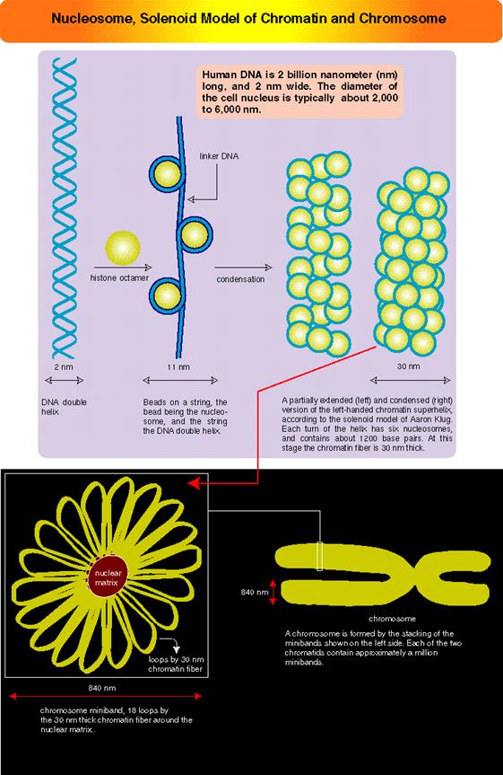
Chromatin: Suspended within the
nuclear sap are the net works of threads, when stained they take color; hence
they are called chromatin threads (chroma-colour, tene-thread). Some of the
threads, particularly their ends are associated with either pore complexes or
inner nuclear membranes. Chromosomal threads are attached to a proteinaceous
matrix at the inner surface of the nuclear membrane, thus the positions of
chromosomal threads is fixed. This interphase chromatin network is not a
constant feature, but changes as and when the cell passes through various
stages of cell division. Nevertheless, the chromatin at the G1 stage, appears
to be diffused, thin, single stranded and coiled, but this single stranded
chromatin threads undergo duplication to form double stranded chromatin threads
at S-stage. During duplication chromosomal DNA replicates, and necessary
histones and nonhistones are drawn from the nuclear sap to form sister
chromatin threads. With the progress of interphase into prophase, the long,
thin threads undergo a continuous process of condensation resulting in shorter
and thicker chromosomes. At the same time, the chromatin distangles from the
network and chromosomes slowly get resolved into individual threads.
By the time, cells
reach metaphase stage, chromosomes undergo maximum condensation, and individual
chromosomes can be made out. At this stage the number and the detailed
structure of them can be studied with light microscope or electron microscope.
Number of
chromosomes: The number of chromosomes varies from organism to
organism (2-1600) and this number is constant and characteristic for a given
species. (Table below).
|
Common
name
|
Specific
name
|
Chromosomal
number(2n)
|
Fruity fly |
Drosophila
|
8
|
|
Frog
|
Rana pipiens
|
20
|
|
Gorilla
|
Gorilla gorilla
|
48
|
|
Monkey
|
Macaca mulatta
|
42
|
|
Man
|
Homo sapiens
|
46
|
|
Garden pea
|
Pisum sativum
|
14
|
|
French bean
|
Phaseolus vulgaris
|
14
|
|
Onion
|
Allium cepa
|
16
|
|
Cabbage
|
Brassica oleracea
|
18
|
|
Coffee
|
Coffea arabica
|
44
|
The number of
chromosomes is denoted by the terms Karyotype which may be either haploid or
polyploid. The haploid karyotype consists of one set of chromosomes, where
every individual chromosome is structurally and genomically different from the
others and exhibit unique characteristics. For example: in the case of onion,
the haploid (n) chromosome number is 8 and let us call them as A, B, C, D, E,
F, G, H. Here, each chromosome is different and unique in its genomic
content. And such a set of chromosomes is called haploid set. If two such sets
of chromosomes are put together in the same nucleus then that nucleus or the
organism that posses it, is called diploid i.e. here two haploid sets are
present, they are called homologous chromosomes or homologous pairs. On the
other hand, as A and B chromosomes are being different, they are called
non-homologous chromosomes. The terms triploid (3 n), tetraploid (4 n) and
polyploid just indicates the number of sets present in the nucleus.
A group of organisms
belonging to a particular species, though show a constant chromosomal number,
say diploid, they often exhibit variation in chromosome numbers, either by loss
or gain of one or more chromosomes. In some cases the entire set of
chromosomes may be involved. This variation in chromosomal number leads to
variation in the morphology and functional behavior of organisms. Such changes
may ultimately lead to variation and origin of species, provided they survive.
This is one of the fundamental steps in organic evolution.
Size of
chromosomes: Chromosomal size varies from organism to organism,
however a particular size of chromosomes is constant for a given species. Some
of the plants of Cyperaceae and Luzula have very small chromosomes, but plants
like Trillium have quite large chromosomes of the size 30µ in length. However,
in a given karyotype, all the chromosomes are not of the same size
(asymmetrical karyotype) and rarely do we find organisms with chromosomes of
the same size (symmetrical karyotype). Some chromosomes like salivary gland
chromosomes (Drosophila), Lampbrush chromosomes, (Xenopus laevis) and
chromosomes in the endosperm haustoria of Phaseolus are 100-1000 times larger
than their somatic chromosomes. These are called special type of chromosomes
or giant chromosomes. They become so because of the necessity. Here each of
the genes is duplicated to thousand times and very helpful in producing
transcripts in large numbers for developmental purpose.
Shape of
the Chromosomes: Almost all chromosomes look like spirally coiled thread,
but during cell division particularly at anaphase stage, chromosomes show a
specific bent shape. This is due to the position of primary constriction or
centromere. Accordingly, the chromosomes are called Metacentric (V-shape), Sub-metacentric
(J-shape), Acrocentric (rod shape),
Telocentric(rod
shape), Polycentric(wave shape) and Diffused (rod shaped but move horizontally).
Sex
chromosomes and Autosomes: Higher organisms like man, monkeys and some plants where
male and female sexes are morphologically differentiated, the cells in them
contain two types of chromosomes, called Autosomes and sex chromosomes. The
latter classification is based on the X and Y chromosomes. This classification
is based on the chromatin nature and function. For example: human beings (Homo
sapiens) have 46 chromosomes of which 44 are autosomes and 2 are sex
chromosomes. Autosomes are believed to control the development of somatic body
and sex chromosomes are responsible for the expression of sexual organs and characters.
If two XX chromosomes are present, it determines the female sex of the organism
and its related characters are expressed; if one X and one Y chromosomes are
present, male character is expressed. The X chromosome is considered to express
female character and Y and the male character. The X chromosomes are more or
less euchromatic and Y are heterochromatic. Furthermore, the sex expression
varies in different organisms. All in all, it is the interaction between autosomes
and sex chromosomes that ultimately determines the expression of sexes through
the mediation of specific hormones like estrogens, female hormones and
Androgens male hormones.
Chromosomal
structure
Using optical
microscopes, if chromosomes of metaphase (at which chromosomes are at maximum
condensation) are observed, chromosomes appear to be double stranded threads,
which are relatively coiled to each other. These threads are referred to as
chromatids or chromonemata. If such chromatids are carefully observed under
high resolution light microscope (2000 times enlarged), each of them appears to
be spirally coiled with apparent gyrations. Many chromosomes show differential
condensation, because of which some parts take more stains and other less stain.
The former is called heterochromatic segments and the later are euchromatic.
This differential staining behavior is called heteropycnosis. The
heterochromatic segments may be either tightly condensed (take more strain) or
less condensed (takes less
stain), this feature is called as positive heteropycnosis and negative
heteropycnosis respectively. In some cases the entire chromosome appears to be
heterochromatic because of greater condensation. The heterochromatin can be
constitutive as in CEN and telomeric regions and it can be facultative also in
other regions; the position of such constitutive regions does not change but
facultative heterochromatin may change during development and from one tissue
to the other. Formerly, these heterochromatic regions were believed to be
genetically inert, now they are known to contain genes and they do express.
However, now, constitutive heterochromatin regions like CEN and telomeres are
know to contain highly repetitive DNA segments.
The inheritance of Mendelian
factors or characters through chromosomes was not substantiated till the
discovery of chromosomes. This has, however, led to unit character
inheritance, specially located within chromosomes. These unit characters are
now called genes. Nevertheless, the problem of genes which are arranged in
chromosomes was an enigma, but the discovery of bead like structures in meiotic
chromosomes, called chromomeres, has given an impetus in unraveling this
problem. Meiotic chromosomes particularly at leptotene stage appear as fine
strings of beads.
These beads like
structures were called as chromomeres and were equated to individual genes.
Such chromomeres were assumed to be held by nongenetic threads. But later,
chromomeres were found be none other than the coiled expressions of chromonemata;
when two ends of such chromatids are stretched apart, the chromomeres
disappear. Nevertheless, the concept of linear arrangement of genes in
chromosomes has been accepted. Morgan and later Hunt’s cytogenetic experiments
have furthered the concept of gene as a unit of heredity, a unit of
recombination, a unit of mutation and a unit of function and chromosomes form
unit of linkage.
Morphologically the
metaphasic chromosomes appear to be simple, coiled threads of uniform
thickness; here and there the chromosomes contain constricted or narrow
regions. These are called primary constrictions and secondary constrictions
respectively. The primary constrictions and secondary constrictions are
further differentiated and characterized by their behavior and functions.
Centromere: The primary
constriction is also called centromere for it is the region at which
chromosomes get attached to tractile fibres and they lead the anaphasic
chromosomal movements. If the centromere is destroyed by direct X-radiation
hits, chromosomes behave abnormally and lose their directional movements. This
results in loss or gain chromosomes at the end of cell divisions. Thus
centromere appears to be a non stainable gap in the chromosomal thread.
However, in metaphasic chromosomes, though the arms are double stranded, the
centromere still appears to be single stranded, but in actuality it is double
stranded but held tightly by proteins called adhesins.
Recent electron microscopic
studies clearly show that on either side of the centromeric region, in line
with the chromosomal arms, it is covered by or ‘C’ shaped structure called
Kinetochore. Thus mitotic chromosomes contain two kinetochore structures are present
at each centromere. Each kinetochore is made up of three regions. The outer
most region is cup shaped structure called commissural cup and it is relatively
thick. On its outer surface large number of processes called corona are
present. Amidst the corona a number of (3-10) microtubules are found to be
penetrated as deep as to reach chromonema. The middle region is less dense,
but the inner region is dense and it is in contact with centromeric
chromonema. However, the entire kinetochore structure appears to be a highly
specialized region and it is likely to participate as the nucleating centre in
the polymerization of tubulin into tractile fibers during the formation of
mitotic apparatus.
Another interesting
feature of the centromere is that, on both flanking regions of the centromere
the chromatin material is heterochromatic. And it is now known that the DNA in
this region are highly repeated (highly repetitive class of DNA), however their
function is not known.
Secondary
constriction or Nucleolar organizer: Similar to that of primary
constriction another constriction is present only on specific chromosomes.
Such constriction is called secondary constriction and it is characterized by
its nucleolar formation, hence it is also referred to as Nucleolar organizer.
This region contains DNA segments for ribosomal RNA. The genes present, here,
are redundant or tandemly repeated. In some cases, like frog oocytes these
genes get amplified at the end of the telophase. The DNA in the region opens
out, and starts transcribing ribosomal RNA as large precursors. Later this RNA
is sliced and processed into 18s 28s and 5.8s RNAs. These RNAs in turn get
associated with different ribo-proteins sequentially and functional ribosomes
are produced. If the secondary constriction segment of the chromosome is
knocked off, the nucleolus fails to appear and the cells die prematurely. In
human chromosomes the nucleolar organizers are found on chromosomes 13, 14, 15,
21 and 22.
SAT
Chromosomes: Short chromosomal segments, at the terminal region contain
blob like structure, beyond secondary constriction; they are called Satellites.
Such chromosomes are called SAT chromosomes. Heitz, who coined this term, is
in the opinion that these segments are lacking in thymidilic acid, i.e. Sine
Acido Thymidine (SAT). Otherwise, this region, as it is lacking Thymidilic
acid, it is rich in Guanidilic and Cytidilic acids. Whether such GC rich
heterochromatic chromosomal segments contain some redundant genes or not, is
not known. They are rich in short tandem repeat DNA segments. Their function
is also a mystery.
Telomeres: In all most all
organisms, with out any exceptions, chromosomal ends have heterochromatin
materials. Such structures are referred to as telomeres. The presence of such
structures are found
to be non sticky and prevent the attachment of broken chromosomal ends in wrong
way. If telomeres are cut off, the ends become sticky. However, the recent in
situ hybridization techniques have demonstrated that heterochromatic telomeres
contain repetitive DNA of particular sequence such as GGGATT. The telomeric
DNA runs many thousands of base pairs of such repeats.
The DNA analysis of
such ends show, the 3’end of the DNA in these regions is free and can be
subjected to exonuclease action. So in order to protect such ends, in this
region the DNA is organized into loop like structure associated with a variety
of proteins to protect from exonucleases. Even the replication of this
telomeric DNA uses a short RNA as the primer and reverse transcriptase for
generating telomeric DNA. However the size of telomeres changes; they may
expand or contract. Damaged telomeric ends can cause or induce cancer.
Ultra
structure of Chromosomes
Observations of
metaphasic chromosomes under optical microscopes of reasonable resolution,
shows them as fine, plectonemally coiled threads. Beyond this, it is difficult
to understand the internal organization of chromosomes. Nevertheless, the
techniques, like whole mount chromosomes combined with biochemistry and electron
microscopy, have revealed that chromosomes are made up of highly folded but
coiled chromonemal threads of 250-300 A0 thickness, with ends visible nowhere.
Biochemical combined
with molecular studies studies has revealed that the chromosomes contain DNA,
histone and non-histone proteins. The complexity of the organization of the
above said components has further compounded with the discovery of various
components of histones and nonhistones. Histones are found to be basic
proteins and different kinds such as H1, H2A, H2B, H3, and H4. These proteins
are rich in lysine and Arginine residues that are the reason they show basic
properties.
The nonhistones
proteins are found to be acidic in nature and 100-120 or more different kinds
have been identified by 2-D gels.
When chromatin is
added to a salt medium, the chromatin threads loosen and spread out into fine
bead like structures called Nu-bodies or Nucleosomes. The Nu-bodies were
further identified as to contain histones as octamer (2H2A 2H2B, 2H3 and 2H4)
and DNA double helix of about 140 base pairs length is coiled around it. The
nucleosomes are the fundament chromonemal units, where each of the histone as
tetramers are superposed one on each. Each of the histones contain a folded
structure with free N-terminal tails and H2A contains a C-terminal tail too.
They play very important role in condensation of the chromonema and
decondensation and differential gene expression. The amino acid sequence in the
tail provides a kind of signatures such can utilized as Chromodomain and
Bromodomain, very very essential gene expression or repression. The DNA thread
found in between two such Nu-bodies is called linker DNA which consists of 50
to 100 base pairs.
Furthermore, the
electron microscopic studies of polytene chromosomes reveal that the Nu-bodies
or nucleosomes are biconvex disc shaped structures with histone octamer as the
core, around which DNA coiled 1 ¾ times. These structures have been recognized
as the fundamental units of chromosomal fibres.
Such fundamental
chromosomal fibres with Nu-bodies as units, undergo solenoid coiling or produce
compact zigzag conformation in the presence of H1 proteins. This forms the
basic thread of chromosomes called chromonema of 30nm size. Each of the
solenoid or zigzag structures may contain about 6-7 nucleosomes. Then this
solenoid thread, in association or in combination with specific proteins,
undergoes further compaction with emanating loops from the scaffold. The DNA
in the loops of different sizes are attached to scaffold proteins by a specific
DNA sequences called MARs, called so they are matrix attachments regions. The size
of the loops vary in their
structure and
dimensions; the dimensions can be of 300A size. These chromonemal compact structures
are visible under light microscope. These threads show a contraction of 1300
to 1500 fold to that of string of beads.
Artificial, these are man made synthetic chromosomes, look
at the arrow pointing to an artificial-synthetic chromosome:
Such chromonemata are
further coiled in metaphasic chromosome which exhibits a 6 x 10³ – 7 x 10³ fold
condensation. However the coiling and condensation of Nu-bead fibres into
microscopically visible chromosome, is aided and augmented by a number of non-histone
proteins as binding factors to form that can be observed under optical
microscopes. If such chromosomes are subjected to histone digestion and
dispersed under certain detergent cum salt solution, the histones are
selectively removed, but retain the non-histone proteins intact. All the
histone free DNA molecules spill out in the form of loops of fibers of various
sizes.
These and other
studies, notwithstanding the complexity of the association of DNA with
proteins, have shown that each chromonemata is made up of single, but a long
DNA molecule, which supports the concept that the genes are arranged in linear
order, where a segment of DNA acts as the unit of heredity. Such chromosomes go
through condensation and relaxation during cell division in stage specific
manner. In interphase most of the chromosomes are relaxed and bound to inner
lamina proteins and found in fixed positions.
In recent years, with
greater understanding of the structure and functions of chromosomes, molecular
biologists have created chromosomes in the lab, they can be stained and
observed along with others normal ones. This will provide great benefits to
cure certain hereditary diseases, I am sure not immediately, it may take few
more years.
Giant chromosomes
Chromosomes, in most
of the organisms show a cycle of condensation and decondensation during various
stages of cell divisions. But in some organisms, chromosomes enlarge
considerably to the tune of 250-500 times the normal somatic chromosomes. The
difference in their sizes can be observed under optical microscopes with lower
resolution. These are called giant chromosomes and they are not just restricted
to one species but found to occur in various organisms like insects, frogs,
salamanders and even plants.
However, their occurrence
is restricted to certain stages in the life cycle. Two such giant chromosomal types’
i.e. salivary gland chromosome and Lampbrush chromosome have been extensively
studied.
Salivary
gland chromosomes or polytene chromosomes: Salivary gland chromosomes
are found in salivary gland cells of 11th day larvae of dipteron class of
insects. They are present in the larvae of Chironema, Drosophila melanogaster,
mosquitoes and other insects of the same class. Such giant chromosomes are
also found in the endospermal haustoria of Phaseolus vulgaris.
In the somatic cells
of the insect, these chromosomes remain normal in their size, but on hatching
of eggs and when the larva reaches the 11th day and is about to undergo
metamorphosis into pupa, the somatic chromosomes found in the salivary glands undergo
a dramatic change in the size and activity. At this stage of development,
chromosomes undergo repeated chromosomal DNA replication without separation,
which leads to the multiplication of two chromonemal threads into 1000-1084
strands.
Furthermore, these
somatic homologous pairs of chromosomes are found to be in pairs or synaptic
state.
Structurally these
chromosomes contain thousands of genes longitudinally oriented and the chromonemal
threads arranged parallel to each other. These threads also show cross banding
of various sizes. Some bands take greater stain and others stain less. The
Darkly stainable bands are heterochromatin segments where the chromosome is
densely packed with chromatin material.
Occasionally some of
these darker bands containing highly condensed chromatin or chromatin in the
inter bands open out into nacked DNA loops and same are transcribed, and one
can find RNAPs associated with transcribed m.RNAs. Even some inter-band regions
also open out. The transcriptional activity has been identified by the use of
radioactive precursors. This region represents intense gene expression
activity. Because of this reason, this region appears be puff like structure
which consists of opened out DNA strands in the form of a ring like structures
called Balbiani rings. In each loci thousands of loops of various sizes can be
seen, so appear as puff like structures. Such puffs can be induced in the
chromosomes by applying an insect hormone called ecdysone. This hormone
induces the transformation of larva into pupa, during which many morphological
structures and functional activities of the larva undergo dramatic changes. Probably,
because of these reasons, the 11th day larval chromosome undergoes differential
gene activation to synthesize required protein products for the metamorphosis
in stepwise manner. During the course development many of the genes expressed
earlier, get silenced and new set of genes get expressed in a form called
cascading, that is one leads to the other, a differential expression required
for the metamorphosis. Many of the genes expressed during these stages have
been identified.

Lampbrush chromosomes: Chromosomes which appear as bottle brush
or the brush that is used to clean the lamp glasses are often called Lampbrush
chromosomes. These chromosomes are highly elongated (5900 um) and they are
found in the oocytes frogs. Here during the development of oocytes the homologous
pairs of chromosomes undergo into extended threads by the uncoiling of specific
chromomere and differentiation. To begin with, the homologous pairs undergo
synapsis, and later they are held to each other at chiasmatic regions. Each
homologous chromosome consists of two chromonemata containing a large number of
granular structures called chromomeres. Many of these chromomeres, all along
the length of chromatin threads open out in the form of large lateral loops of
nacked DNA. Soon, these loops will be covered by a matrix, which is made up of
a pool of RNAs and RNA polymerases.

These lateral loops
now start intense transcriptional activity, with the result, innumerable RNAs
are synthesized. Each loop consists of many genes of the same kind or
different separated by noncoding regions called spacer DNA.
Transcriptional
activity is initiated at several sites simultaneously by the binding of RNA
polymerases and soon RNA strands are formed with the progression of RNA
polymerase. As soon as the initiating site is free, another RNA polymerase
settles and starts synthesis at various stages and each of these segments
appears as the branch of a christmas tree. RNA synthesis is required for the developing
egg and many of the poly A-RNAs (mRNAs) are stored to be translated only when
the egg is fertilized. This is another instance of differential gene activity,
required for certain development process.
No comments:
Post a Comment